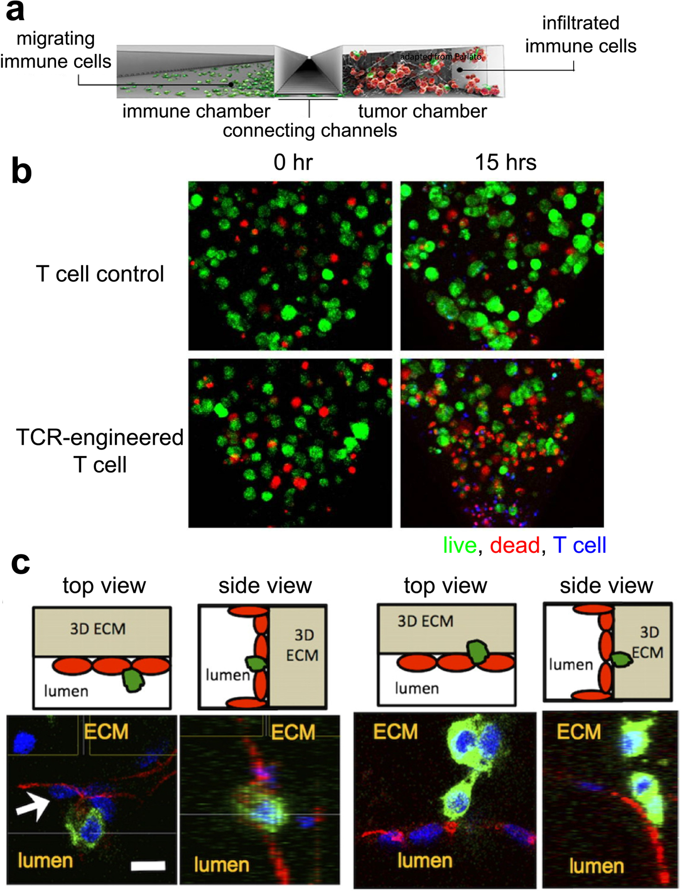
Figure 4.

(a) A side section of a typical microfluidics device. A channel connects the immune and tumor chamber, allowing for crosstalk between the two cell types. (b) TCR-engineered T cells (blue) showed tumor (green)-specific cytotoxicity (red) over time, compared to control T cells (ref [151]). (c) Endothelial cells (red) lining the surfaces of a microfluidics channel interacted with tumor cells (green) extravasating (ref [143]).
