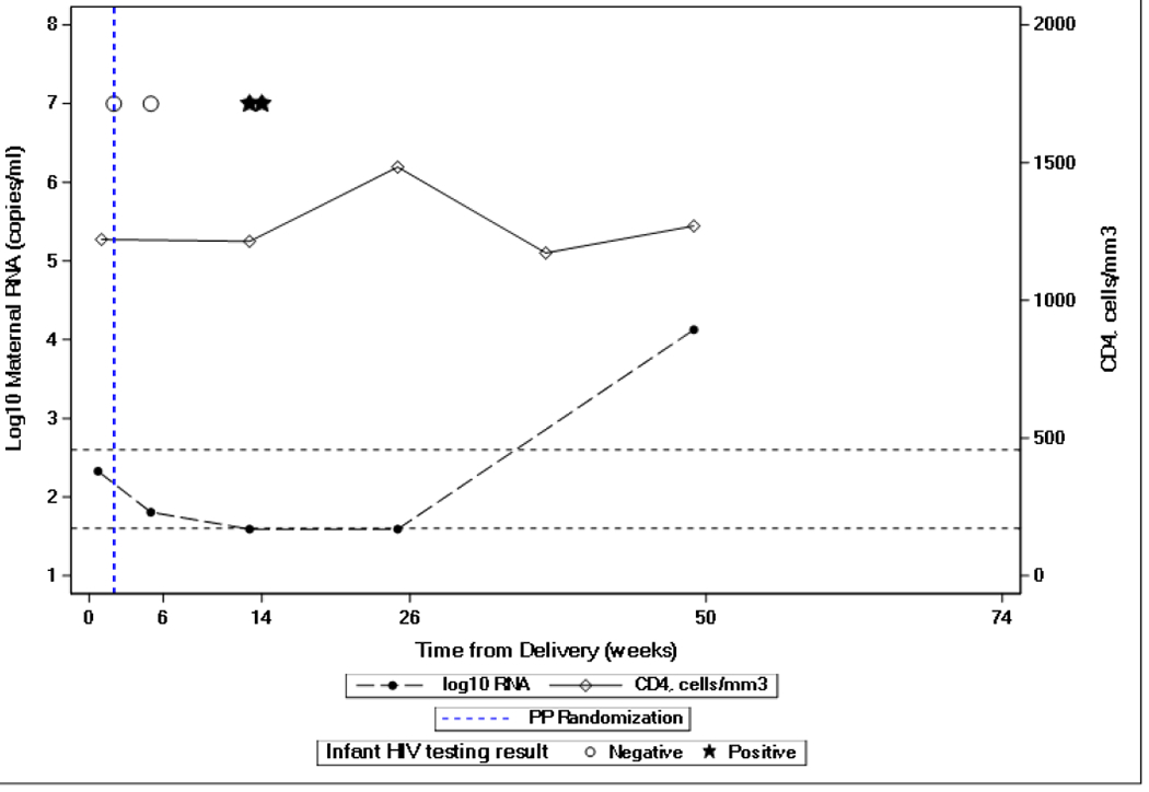
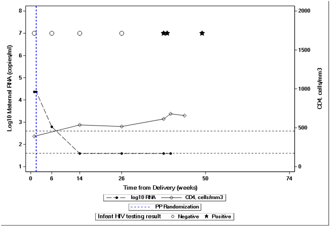
Figure 1.


Maternal plasma HIV-1 viral load and infant NAT testing of two infants with maternal HIV-1 viral load “non-detected” or “detected but below 40 copies/mL” prior to positive infant HIV-1 NAT testing. Dashed lines show maternal HIV-1 viral load results. Solid lines show maternal CD4 cell count. Open circles represent negative infant HIV-1 NATs and black stars represent positive infant HIV-1 NAT.
