
FIGURE 5:
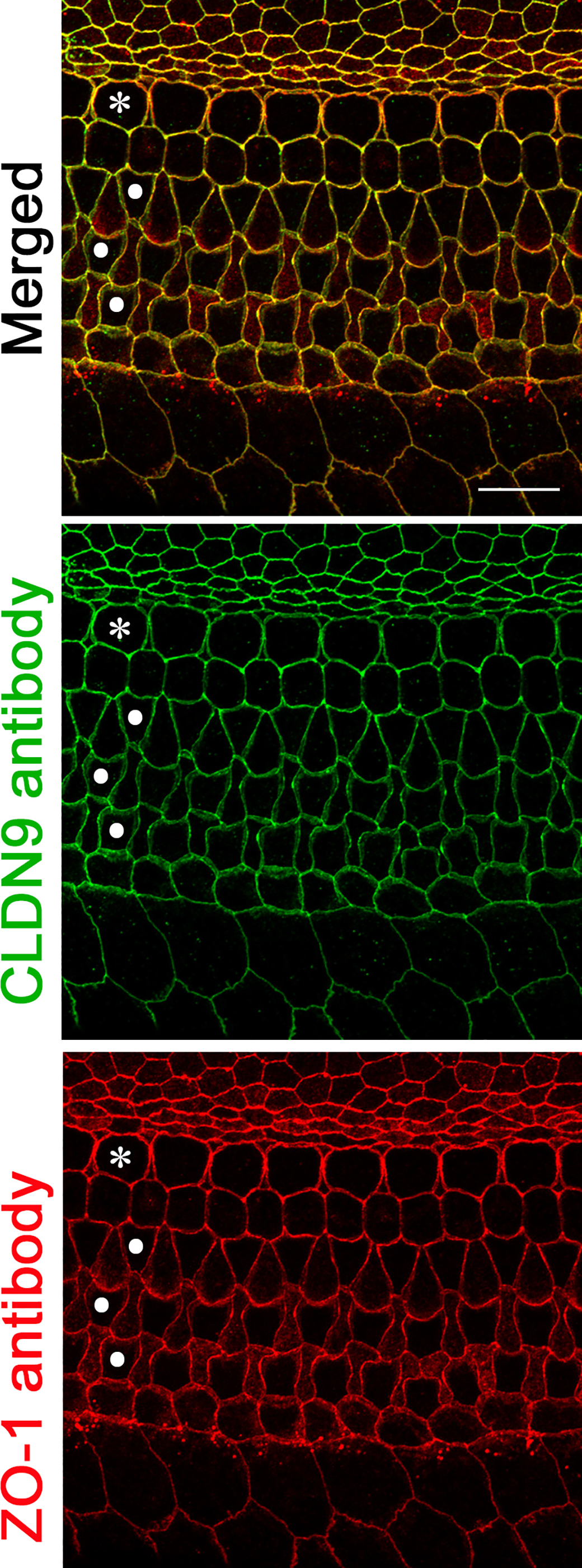
Localization of CLDN9 in mouse inner ear detected by immunofluorescence confocal microscopy. CLDN9 is present in tight junctions of P6 C57BL/6J mouse outer and inner hair cells as well as in all non-sensory cells outlining scala media, including marginal cells of stria vascularis and the epithelial cells of the Reissner’s membrane. CLDN9 antibody labeling is shown in green, anti-ZO-1 antibody labeling is in red. Yellow indicates a merged signal from both antibodies, indicating the co-localization of the two detected proteins. Scale bar is 10 μm. * indicates outer hair cells and • indicates rows of inner hair cells.