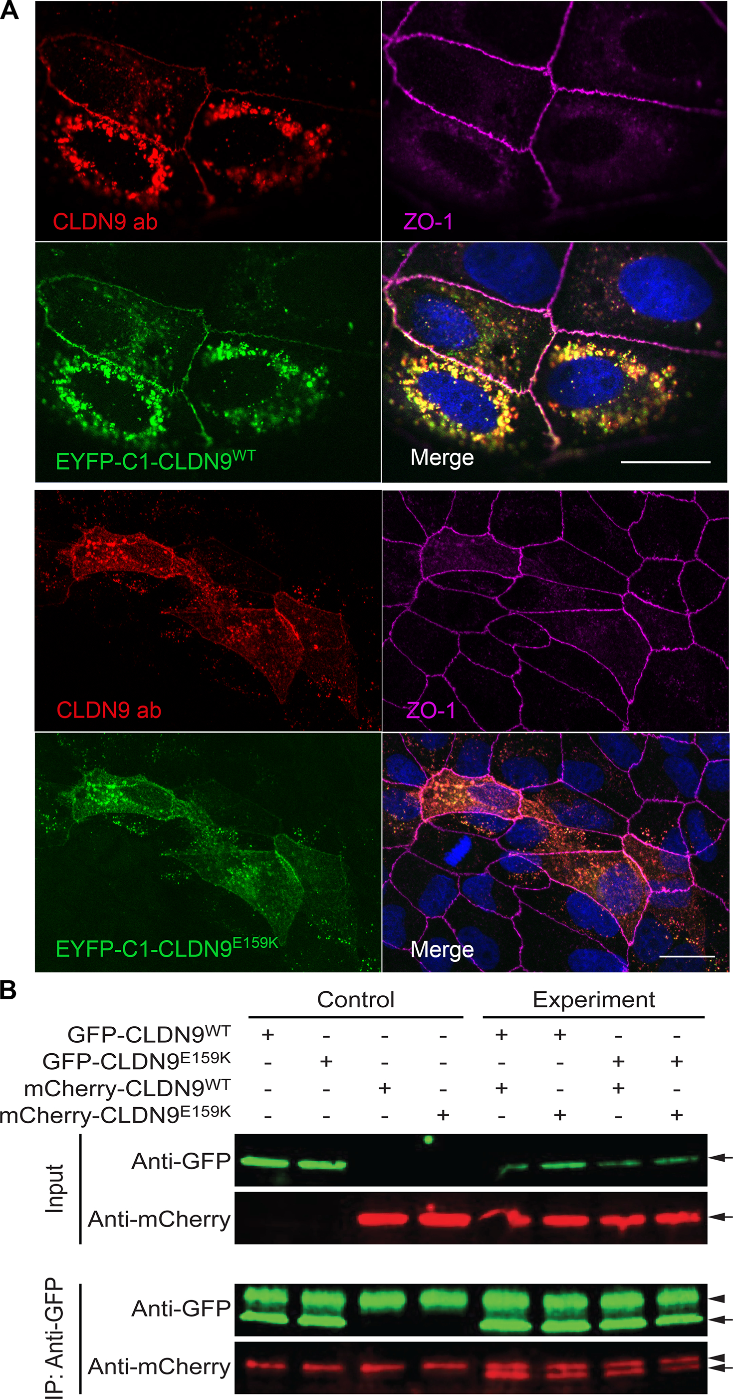
FIGURE 6:

Localization of overexpressed wild-type and mutant CLDN9 in MDCK-II cells and a pulldown assay showing that cis-interaction of CLDN9 molecules is not affected by the Glu159Lys variant. A, Maximum intensity projection images for localization of CLDN9 in MDCK-II cells. Both wild-type (EYFP-C1-CLDN9WT) and mutant constructs (EYFP-C1-CLDN9E159K (Glu159Lys)) were targeted to the cell membrane and were present in the regions of cell-to-cell contact. ZO-1, a tight junction associated protein in the cell membrane (indicated in magenta), was used as a tight junction marker. Anti-CLDN9 antibody (CLDN9 ab, red) was used as an additional marker to detect EYFP-C1-CLDN9WT and EYFP-C1-CLDN9E159K (depicted in green). The localization of CLDN9 was not affected by Glu159Lys substitution as shown by the comparison of merged images (top and bottom panels). Scale bar is 20 μm in both panels. B, Lysates from HeLa cells transfected with both CLDN9WT and CLDN9E159K (Glu159Lys) tagged with EGFP or m-Cherry expression constructs in various combinations were used for co-IP assays with anti-GFP antibody-coated beads. Precipitates were immunoblotted with antibodies against EGFP and m-Cherry, expected sized bands were observed (arrows), Antibody heavy chain bands were also observed in all the pulldown sample lanes (arrowhead). CLDN9WT was able to pull down both CLDN9WT as well as CLDN9E159K. In addition, CLDN9E159K was also able to pull down CLDN9E159K, indicating that Glu159Lys change does not affect the claudin 9 cis-interaction.
