. 2021 Sep 15;22(9):774–781. doi: 10.1631/jzus.B2000793

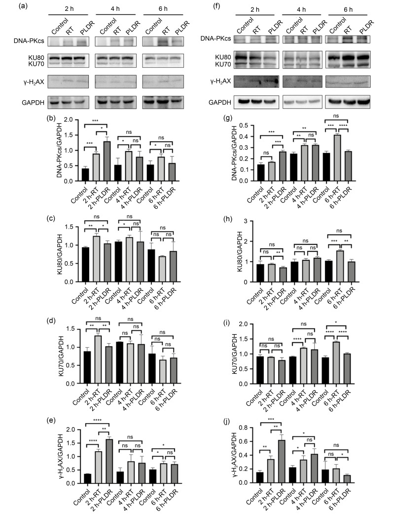
Fig. 5. Effects of RT and PLDR schedules on the expression levels of DNA damage proteins in PANC-1 (a‒e) and PC3 (f‒j) by western blot. PANC-1 (a) and PC3 (f) cell lysates were collected at different time points for western blot. The band intensities of DNA-PKcs (b, g), KU80 (c, h), KU70 (d, i), and γ-H2AX (e, j) in PANC-1 (b‒e) and PC3 (g‒j) cells were quantified after normalized to GAPDH. * P<0.05, ** P<0.01, *** P<0.001, **** P<0.0001. Values are presented as mean±SD (n=3). RT: radiotherapy; PLDR: pulsed low-dose rate; DNA-PKcs: DNA-dependent protein kinase catalytic subunit; γ-H2AX: phosphorylation of H2AX at Ser139; GAPDH: glyceraldehyde-3-phosphate dehydrogenase; SD: standard deviation; ns: not significant.

Fig. 5