
FIGURE 1.
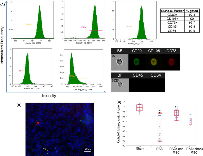
MSCs surface markers, tracking and their effects on the kidney size in mice with renal artery stenosis. (A) Flow cytometry analysis and representative cell images show that human adipose tissue‐derived mesenchymal stem/stromal cells (MSCs) are positive for CD73, CD90 and CD105, but negative for CD45 and CD34. (B) Representative image showing no co‐localization of MSCs (red arrow) and CD3+ T lymphocytes (green arrow) in the stenotic mouse kidney. (C) The ratio of the stenotic and contralateral kidney weights was decreased in mice with renal artery stenosis (RAS) compared to sham and improved in mice injected with lean MSCs, but not with obese MSCs. *p < 0.05 vs. sham, # p < 0.05 vs. RAS