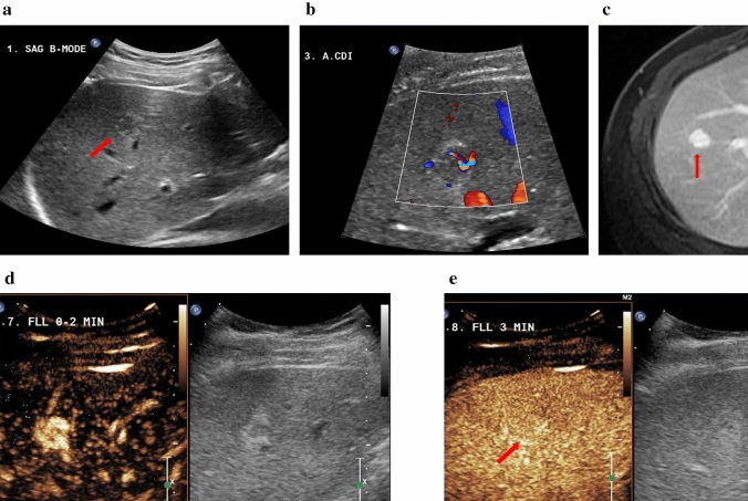
Fig. 4.
34-year-old male with focal nodular hyperplasia (FNH). a Pre-contrast harmonic B-mode showing the lesion. b In Colour Doppler imaging the lesion shows central vascularity and some peripheral vascularity. c The hyperintense representation of the lesion in the venous/delayed phase on CE-MRI. Imaging performed post-SonoVue injection. d Arterial phase shows well-defined lesion margins and central scarring. e FNH homogeneous iso-enhancement indicated in the late phase at 3 min

