Fig. 10.
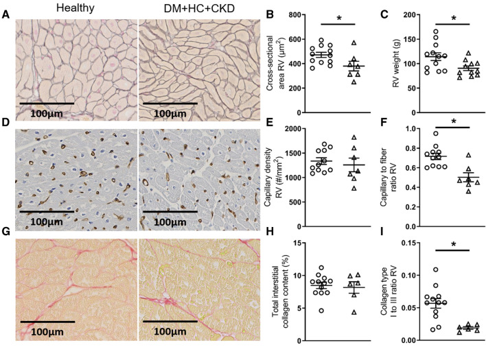
Right ventricular structure of DM + HC + CKD and Healthy swine typical examples of right ventricular Gomori stained (A), Lectin stained (D) and Picrosirius red stained (G) sections. The cross-sectional area of the RV cardiomyocytes was decreased in DM + HC + CKD swine (B), RV weight was also lower in DM + HC + CKD swine (C). Capillary density was similar between groups (E) but capillary-to-fiber ratio was lower in DM + HC + CKD (F). Total interstitial collagen content (H) was unaltered in DM + HC + CKD swine, but there was a shift the composition of the specific collagen fibers in DM + HC + CKD (I). Values are mean ± SEM. *P ≤ 0.05 for Healthy versus DM + HC + CKD, scale bars are 100 µm

