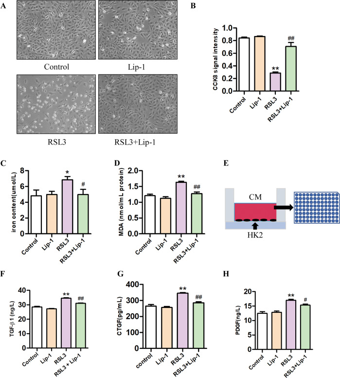
Fig. 5. Effects of Liproxstatin-1 on RSL3-induced HK2 cells ferroptosis and profibrotic factor secretion.
A The morphological of HK2 cells by a light microscope after the treatment for 24 h. B Viability of HK2 cells, evaluated by the CCK-8 assay (n = 3, **P < 0.01 vs control group; ##P < 0.01 vs RSL3 group). C Iron concentrations in HK2 cells (n = 6, *P < 0.05 vs control group; #P < 0.05 vs RSL3 group). D MDA levels in the HK2 cells (n = 6, **P < 0.01 vs control group; ##P < 0.01 vs RSL3 group). E Experimental design. F–H TGF-β1, CTGF, and PDGF levels in the HK2 cells conditioned medium (n = 3, **P < 0.01 vs control group; #P < 0.05, ##P < 0.01 vs RSL3 group). The data are presented as the mean ± SD.

