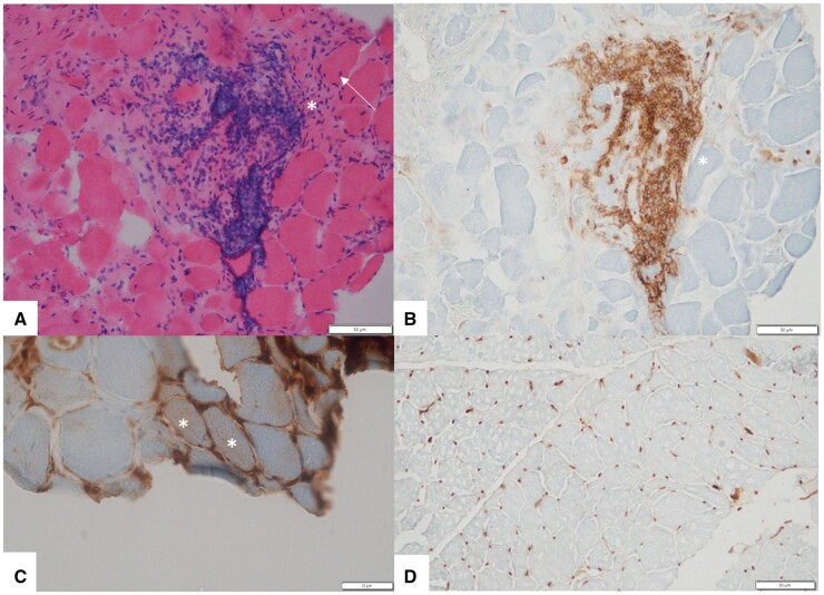
Fig. 1.
Myopathologic features in this patient
(A) Haematoxylin and eosin–stained section shows variation in fibre size, few internal nuclei (arrow) and a large perimysial inflammatory infiltrate (asterisk). (B) CD45-stained section highlights CD45-positive perimysial inflammatory cells (asterisk). (C) MHC-1-stained section shows two fibres with sarcolemmal staining (asterisks). (D) MHC-1-stained control section shows normal capillary staining and no sarcolemmal upregulation.

