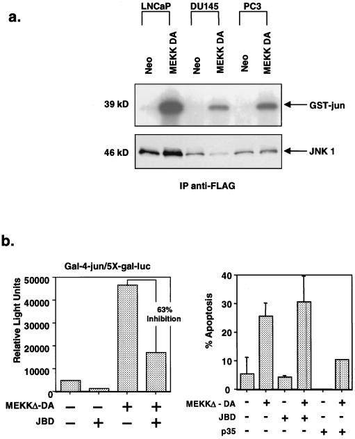
FIG. 4.
Role of JNK activation in MEKKΔ-DA-induced apoptosis. (a) Comparison of JNK activation in prostate cancer cell lines in response to MEKKΔ-DA. LNCaP, DU145, and PC3 cells were transfected with FLAG-tagged JNK1 (2 μg) and cotransfected with Neo or MEKKΔ-DA (2 μg). The top panel shows a JNK assay in which 100 μg of total cellular protein was immunoprecipitated with anti-FLAG antibody and reacted with GST–c-jun. The bottom panel shows an anti-FLAG immunoblot. LNCaP cells have approximately sixfold-higher amount of transfected JNK1 than DU145 cells as determined by densitometry analysis. When corrected for this difference in transfected protein, MEKKΔ-DA-induced JNK activation is approximately four- to sixfold in all three cell lines. (b) Effect of JNK inhibition on MEKKΔ-DA-induced apoptosis in LNCaP cells. LNCaP cells were cotransfected with MEKKΔ-DA (0.6 μg) or Neo and pCDNA3-JBD (JNK1 inhibitor), p35 (caspase inhibitor), or vector control. (Left panel) Effect of transfected JBD on MEKKΔ-DA-induced c-jun transcriptional activity as measured by a 5X-Gal-luciferase reporter (0.4 μg) and gal4-jun (0.4 μg). (Right panel) Transfected cells were scored for apoptosis 48 h after transfection.

