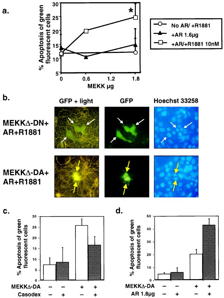
FIG. 5.
Modulation of androgen receptor function alters the sensitivity of prostate cancer cells to MEKKΔ-DA-induced apoptosis. Reconstitution of the androgen receptor signaling pathway in DU145 cells. DU145 cells were cotransfected with MEKKΔ-DA, as indicated, and androgen receptor (AR) (1.8 μg) in the presence or absence of androgen R1881 (10 nM). This graph is an average of the results from eight independent experiments. The P value for the combined experiments is 0.002 as determined by the paired Student t test for MEKKΔ-DA plus androgen receptor plus R1881 versus MEKKΔ-DA plus androgen receptor. (b) Morphology of DU145 reconstituted with androgen receptor and androgen R1881 and cotransfected with MEKKΔ-DN (top row) or MEKKΔ-DA (bottom row) 48 h after transfection. White arrows indicate GFP-positive cells, and yellow arrows indicate GFP-positive cells showing chromatin condensation. (c) Effect of the androgen receptor antagonist Casodex on MEKKΔ-DA-induced apoptosis. Graph of LNCaP transfected with 0.6 μg of MEKKΔ-DA or Neo control vector and treated with the androgen receptor antagonist Casodex (10 μM) as indicated. Graph represents results of four independent experiments in which 200 green fluorescent cells were counted and scored for cytoplasmic blebbing; P = 0.004 as determined by the paired Student t test for MEKKΔ-DA versus MEKKΔ-DA plus Casodex. Cells were scored for apoptosis at 48 h after transfection. (d) Graph of LNCaP transfected with 0.6 μg of pCDNA3 containing MEKKΔ-DA or the empty vector and cotransfected with androgen receptor (1.8 μg) as indicated. Graph represents three independent experiments in which 200 green fluorescent cells were counted and scored for cytoplasmic blebbing at 48 h after transfection; P = 0.01 as determined by the paired Student t test for MEKKΔ-DA versus MEKKΔ-DA plus androgen receptor.

