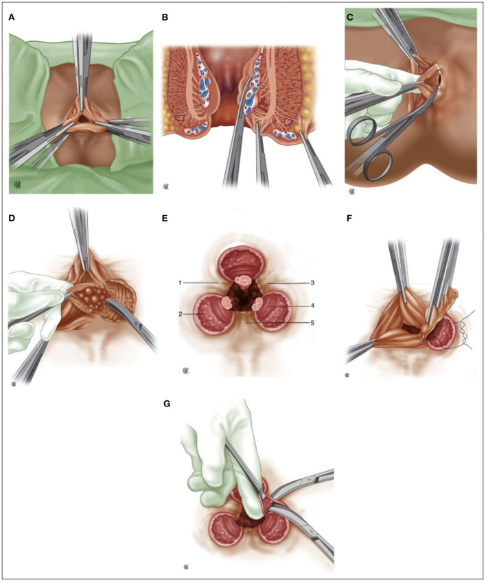
Figure 2.
(A) Milligan-Morgan hemorrhoidectomy, position of the three sets of clamps; (B) Placement of three clamps on the hemorrhoidal group, frontal view; (C) Dissection of the left hemorrhoidal group; (D) Exposure of the internal sphincter after division of Parks' ligament; (E) Final post-operative appearance. 1: left anterior muco-cutaneous bridge. 2: internal anal sphincter. 3: posterior muco-cutaneous bridge. 4: sub-cutaneous fibers of the external anal sphincter. 5: right anterior muco-cutaneous bridge; (F) Suture ligature of the hemorrhoidal pedicle; (G) Cleaning up the muco-cutaneous bridges. (Reproduced from Moult HP, Aubert M, De Parades V. Classical treatment of hemorrhoids. J Visc Surg. (2015) 152:S3–9. Copyright © 2014 Elsevier Masson SAS. All rights reserved).

