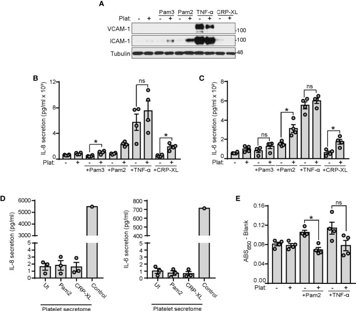
Figure 5.
Platelets enhance endothelial cell inflammation and reduce endothelial permeability to BSA in response to TLR2 agonists. (A–C) HUVECs were cultured onto 12-well plates until reaching confluence and stimulated with Pam2CSK4 (10 µg/ml), Pam3CSK4 (10 µg/ml) or TNF-α (1 ng/ml). 250 µl of platelets (2x10⁸/ml) were mixed with 250 µl of SFM and co-cultured with HUVECs for 4 hours. (A) Adhered platelets were thoroughly removed with SFM and HUVECs lysates analyzed for ICAM-1 and VCAM-1 expression by immunoblot; n=4 experiments. (B, C) Supernatants were collected and assayed by ELISA for IL-6 and IL-8 secretion; n=4 experiments. (D) Washed platelets (1x10⁹/ml) were stimulated with Pam2CSK4 or CRP-XL at 10 µg/ml for 45 min at 37°C. Platelets were then pelleted and supernatants assayed for IL-6 and IL-8 secretion. A positive control from TNF-α stimulated HUVECs was used (Control); n=3. (E) HUVECs were seeded to confluence in transwell devices and stimulated for 4 h at 37°C with Pam2CSK4 (10 µg/ml) or TNF-α (1 ng/ml) in the presence or absence of platelets (1x10⁸/ml). Cells were washed 3 times and incubated with media containing Evans Blue dye (0.67 mg/ml) and 4% BSA for 30 min at 37°C. Samples from the lower chamber were collected and absorbance at 650 nm measured; n=4. * indicates statistical significance (p<0.05). ns indicates not significant. Plat indicates platelets. Error bars indicate standard error of the mean (SEM).

