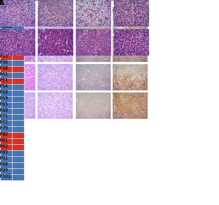
Figure 3.
A comparison of histologic and molecular features between primary (F0) and PDX tumors. (A) Tumor section slides were stained using HE for comparing histology of four F1 PDX tumors with their corresponding F0 tumor; magnification 200×; (B) Consistency of CK19 expression between F0 and F1 PDX tumors. Blue block, CK19-negative expression; red block, CK19-positive expression; (C) Two representative patient samples showing retained pathology and antibody (CK19) status as xenografts over several passages (F1−F3); magnification 200×. HE, hematoxylin and eosin; PDX, patient-derived xenograft; CK19, cytokeratin 19.

