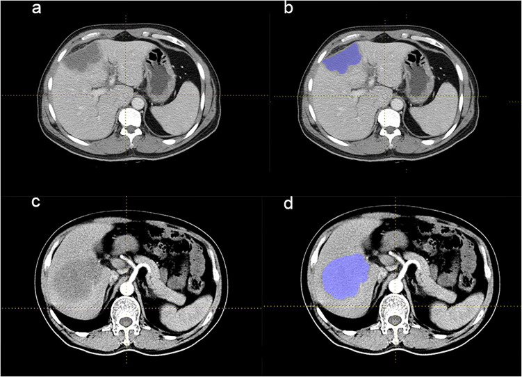
Figure 1.
CECT of patients with ICCA and HL. (a and b) The CECT images of 1 ICCA patient. This patient presented with intermittent dull pain in the right upper abdomen with postprandial pain that had been evident for 8 years and worsened for 20 days. No nausea, vomiting, or yellowish skin staining was found. An irregular and mixed low-density mass was seen in the left internal lobe of the liver, with a blurred boundary and a size of about 8.1 × 3.9 cm on CECT. (c and d) The CECT images of 1 HL patient. This patient was admitted to the hospital for 3 months with intermittent fever, night sweats, and pain in the right chest, accompanied by decreased appetite and no yellowing of the skin. On CECT, a soft tissue mass with slightly lower density was seen in the lower segment of the right lobe of liver, about 8.8 × 8.7 cm, with an ill-defined boundary and uneven moderate enhancement. The boundary between the lesion and the right anterior and posterior portal vein branches was not clear. ROIs were all drawn along the liver lesion slice by slice, and all areas of calcification and necrosis were excluded.
Abbreviations: ICCA, intrahepatic cholangiocarcinoma; CECT, contrast-enhanced computer tomography; HL, hepatic lymphoma; ROI, region of interest.

