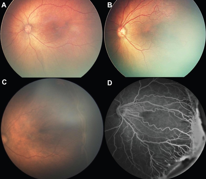
Figure 1.
Clinical manifestation of normal premature infants and those with retinopathy of prematurity who require treatment. (A) Fundoscopy of a premature infant in the control group; (B) Fundoscopy of aggressive posterior ROP: the retina is only vascularized within Zone 1, with apparent tortuosity of retinal arteries and dilation of veins (plus disease); (C) Fundoscopy of stage 3 ROP with plus disease in posterior Zone 2; (D) Fundus fluorescein of ROP, in which prominent retinal neovascularization, plus disease and dragged disc are noted.

