FIG. 1.
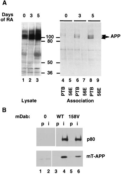
Association between Dab1 and APP. (A) Retinoic acid treatment of P19 EC cells induces their differentiation into postmitotic neurons and glia and induces increased expression of APP as detected by Western blotting of total cell lysates (lanes 1 through 3; days of retinoic acid treatment are indicated above). Cell lysates were incubated with a GST fusion protein containing the wild-type Dab1 PTB domain (PTB; lanes 4, 6, and 8) or a fusion protein containing the 56E mutant PTB domain (lanes 5, 7, and 9). Bound APP was detected by Western blotting. The values between the gels are molecular sizes in kilodaltons. (B) Vector control (lanes 1 and 2) and wild-type (WT; lanes 3 and 4) and 158V mutant (lanes 5 and 6) Dab1 p80 expressed in 293T cells together with the Myc-tagged APP cytoplasmic domain (mT-APP). Immunoprecipitates were prepared by using preimmune serum (p; lanes 1, 3, and 5) or an anti-Dab1 (B3) antibody (i; lanes 2, 4, and 6) and analyzed by SDS-PAGE and Western blotting with antibodies to Dab1 (upper) or the Myc tag (lower).

