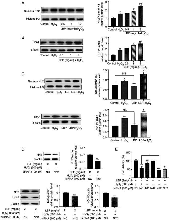
Figure 4.
LBP alleviates H2O2-induced RPE cell damage via the Nrf2/HO-1 pathway. ARPE-19 cells were incubated in the presence or absence of LBP for 24 h, and subsequently treated with 500 µM H2O2 for 2 h. (A) The relative protein expression levels of nuclear Nrf2 were determined via western blotting. (B) The relative protein expression levels of HO-1 were determined via western blotting. (C) Western blot analysis was performed to detect the protein expression levels of nuclear Nrf2 and HO-1. (D) ARPE-19 cells were transfected with siRNA (NC or Nrf2) for 12 h. Protein expression levels of Nrf2 were analyzed via western blotting. ARPE-19 cells were transfected with siRNA (NC or Nrf2) for 12 h, incubated with LBP for 24 h and subsequently treated with H2O2 for 2 h. Protein expression levels of Nrf2 and HO-1 were analyzed via western blotting. (E) ARPE-19 cells were transfected with siRNA (NC or Nrf2) for 12 h, incubated in the presence or absence of LBP for 24 h and subsequently treated with H2O2 for 2 h. The cytoprotective effect of LBP was analyzed via the Cell Counting Kit-8 assay. +, presence of H2O2 or LBP; -, absence of H2O2 or LBP. *P<0.05, **P<0.01, ***P<0.001 vs. control; #P<0.01, ##P<0.001 vs. H2O2-treated cells with no LBP pretreatment. LBP, Lycium barbarum polysaccharide; RPE, retinal pigment epithelium; Nrf2, nuclear factor erythroid 2-related factor 2; HO-1, heme oxygenase-1; si, small interfering; NC, negative control.

