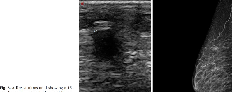
Fig. 3.
a Breast ultrasound showing a 15-mm hypoechogenic solid lesion of the super-external quadrant compatible with the secondary lesion of previous breast cancer. b Diagnostic mammography of the right breast. A well-defined contrast area, with a diameter of about 15 mm of the super-exterior quadrant and in the external para-areolar, 8.5 mm.

