FIGURE 2.

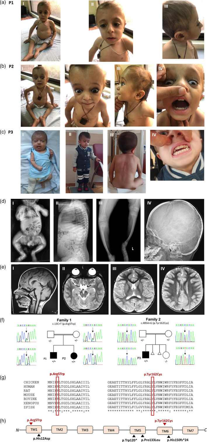
Three affected patients with KDELR2‐related osteogenesis imperfecta from two consanguineous families. (a) Photographs of patient P1 showing short stature, barrel shaped chest (I), sunken eyes, epicanthus inversus (II), and sparse thin hair (III). (b) Photographs of P2 showing short stature, barrel shaped chest (I), blue sclera (II), sunken eyes secondary to molding of the soft cranium (II), thin sparse hair (III), and dentinogenesis imperfecta (IV). (c) Photographs of P3 showing infantile short stature a right leg cast following a pathological femoral fracture (I), current short stature at age 4 years (II), scoliosis (III), and dentinogenesis imperfecta (IV). (d) Radiographs of affected subjects depicting infantile femoral fracture from P3 (I), vertebral compression fractures and platyspondyly from patient P1 (II), short bowed limbs from P1 (III), and Wormian bones from P1 (IV). (e) Brain MRI sections from P1 obtained at 6 years of age. (I) Sagittal T1 showing normal brain appearance. (II) Axial T2 showing brachycephaly. (III and IV) Axial T2 images showing age‐appropriate myelination. (f) Sanger segregation of KDELR2 variants in family 1 and 2. (g) Conservation of amino acid residues across species for both variants. (h) Location of current (red) and previously reported (black) KDELR2 pathogenic variants. All identified variants to date affect transmembrane domains (TMs) 1, 5, and 6 of the KDELR2 protein product [Color figure can be viewed at wileyonlinelibrary.com]
