
FIGURE 2.
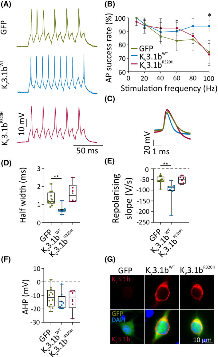
KV3.1bR320H cannot sustain high‐frequency interneuron firing. (A) Representative traces of interneuronal firing at 14–16 days in vitro (DIV) expressing green fluorescent protein (GFP) only, KV3.1bWT, or KV3.1bR320H (100 Hz, 5 ‐ms). (B) Action potential (AP) success rate when stimulated at different frequencies (100 Hz: KV3.1bWT vs. GFP: *p = .04, two‐way repeated measures analysis of variance [ANOVA] followed by Bonferroni multiple comparison test). (C) Representative AP waveforms of interneurons at 14–16 DIV expressing GFP only or KV3.1b channel variants. (D, E) AP half‐width (**p = .004) and the rate of AP repolarization (**p = .005) of KV3.1bWT neurons compared to GFP control neurons (one‐way ANOVA with Bonferroni multiple comparison test). (F) Afterhyperpolarization (AHP) of the first AP. (G) Representative confocal images of lentivirally transduced interneurons at 14 DIV, immunolabeled for KV3.1b. DAPI, 4′,6‐diamidino‐2‐phenylindole (nuclear stain); WT, wild type