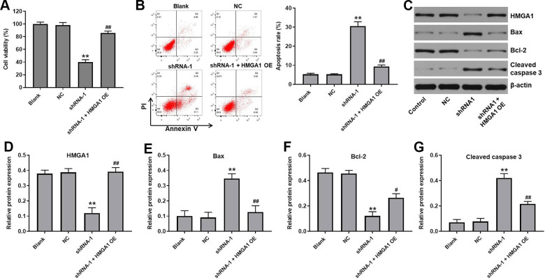
Figure 5.
HMGA1 OE reversed the effect of hsa_circ_0091994 knockdown on the cell viability and apoptosis. AGS cells were administrated with shRNA-1 and HMGA1 OE for 48 h. (A) Cell viability was detected using CCK-8 assay. (B) Cell apoptosis was detected using Annexin V and PI double staining; cell apoptosis rate was quantified. (C) The protein expression of HMGA1, Bax, Bcl-2, cleaved caspase 3 was examined using western blot assay. β-actin was used as a loading control. (D–G) The protein expressions of HMGA1, Bax, Bcl-2, and cleaved caspase 3 were quantified respectively. **P<0.01, compared with blank; n = 3.

