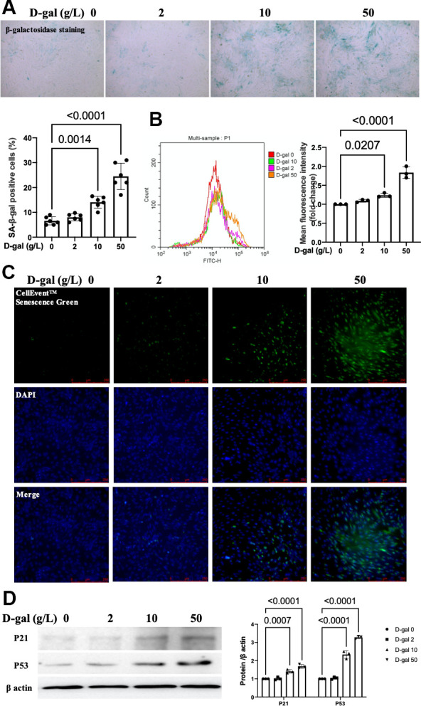
Figure 1.

D-gal induced-H9c2 cells caused cardiocytes aging changes in a concentration-dependent manner. H9c2 cells were treated with different concentrations of D-gal (0, 2, 10 and 50 g/L) for 24 hours. (A) The H9c2 cells senescence induced by D-gal was identified by β-galactosidase staining (100×). Representative bright-field photomicrographs were captured. The blue-stained cells were designated as aging cardiocytes. The blue-stained cells and the total number of cells were counted, and the percentage of cells expressing β-galactosidase was calculated. (B) Flow cytometry analysis was applied to detect the β-galactosidase mean fluorescence intensity after different D-gal concentration senescence induction. (C) H9c2 cells were stained using the CellEvent™ Senescence Green Probe (200×). An increase in senescence-associated β-galactosidase expression, a hallmark for the onset of senescence, could be detected by the CellEvent™ Senescence Green Probe with a fluorescence microscope. (D) The aging-associated proteins (P53, P21) were detected by western blot, and the corresponding quantification was present.
