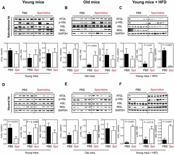
Figure 2.
Spd on lipolysis of white adipose tissues. Western blots for the activity of lipolysis, indicated by ATGL, phosphorylation of HSL S563 (p-HSL), and MGL, in subcutaneous fat of (A) young mice, (B) old mice, and (C) young mice under HFD after 6 mo of Spd treatment. Relative ATGL levels (normalized to GAPDH), p-HSL levels (normalized to HSL), and MGL levels (normalized to GAPDH) were quantified underneath. Activity of lipolysis in the other white adipose tissue, visceral fat, of (D) young mice, (E) old mice, and (F) young mice under HFD. All bars represent mean ± SEM. Significant differences were taken when p <.05. ATGL = adipose triglyceride lipase; GAPDH = glyceraldehyde 3-phosphate dehydrogenase; HFD = high-fat diet; HSL = hormone-sensitive lipase; MGL = monoacylglycerol lipase; PBS = phosphate-buffered saline; Spd = spermidine.

