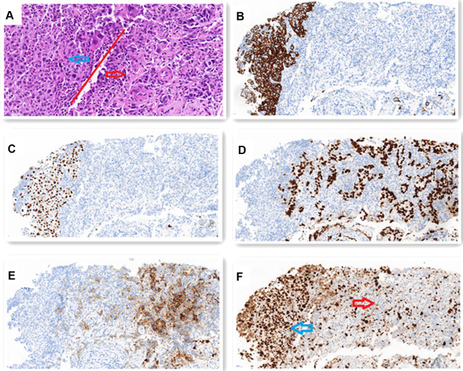
Figure 2.
(A) H-E staining shows that SCC (blue arrow) presents a solid sheet-like arrangement. The cell cytoplasm is red stained, the nucleus is large, nucleoli and mitosis, no intracellular keratinizing and keratinizing beads are seen, but AC (red arrow) is adenoid, cord-like or solid arrangement. The cell cytoplasm is reddish or translucent, the nucleus is large and irregular, and nucleoli and mitosis can be seen. (x200). SCC accounts for about 10% and AC accounts for about 90% of the tumor. IHC staining shows: (B) CK5/6 and (C) P40 are positive in SCC components. (x200) (D) TTF-1 and (E) Napsin-A are positive in AC components. (x200) (F) Ki-67 index is about 70% in SCC (blue arrow) and about 30% in AC (red arrow). (x200).

