FIG. 1.
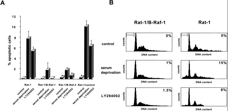
Induction of apoptosis by growth factor deprivation and PI 3-kinase inhibition. (A) Wild-type Rat-1 cells (Rat-1), transfected Rat-1 cells overexpressing B-Raf (Rat-1/B-Raf-1 and -2), and transfected Rat-1 cells that fail to overexpress B-Raf (Rat-1/control) were maintained in normal growth medium with or without the addition of 50 μM LY294002 or were incubated in serum-free medium for 16 h. Cells were then fixed, permeabilized, and stained with the DNA dye bisbenzimide (Hoechst 33258), and the apoptotic nuclei were scored on the basis of nuclear morphology. Data were averaged from three experiments, and at least 300 cells were counted per experiment. The error bars are standard errors of the mean. (B) Rat-1/B-Raf-1 and Rat-1 cells were maintained and treated as described in the legend to Fig. 1A. They were then collected and stained with propidium iodide, and the DNA content was analyzed by flow cytometry. The bar indicates cells with sub-G1 DNA content, a characteristic of apoptosis. The percentage of cells with sub-G1 DNA content is indicated in the upper right corner of each panel. The results are representative of at least two similar experiments.

