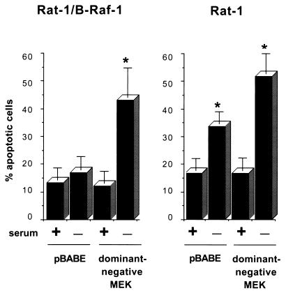
FIG. 4.
Induction of apoptosis by ectopic expression of dominant-negative MEK. Cells were cotransfected with an expression vector for dominant-negative MEK or an empty vector control (pBABE), together with an expression construct for GFP. Cells were maintained in normal growth medium or in serum-free medium for an additional 24 h. Transfected cells were then identified by fluorescence microscopy and scored for apoptosis on the basis of nuclear morphology. Data are presented as the percentage of GFP-positive cells with apoptotic nuclei. Data were averaged from three experiments, and 100 cells transfected with each vector were counted per experiment. The error bars are standard errors of the mean. Asterisks indicate a significant difference (P < 0.05) from controls maintained in serum (analysis of variance test).

