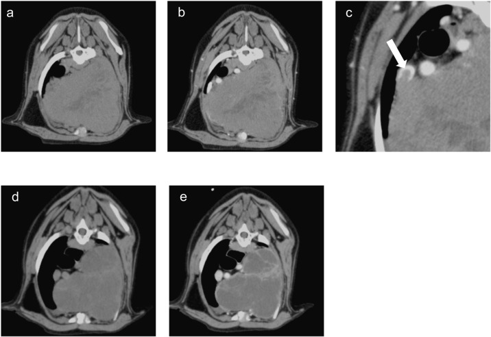
Fig. 3.
Transverse computed tomography (CT) images of the thorax on day 29 (before radiotherapy [RT]; a, b, and c) and 43 (last day of RT; d and e). a and d. plain CT axial view. b, c, and e. CT axial view in the portal venous phase. There was a mass with pale contrast enhancement in the cranial mediastinum (a and b), and tumor invasion into the vena cava indicated by white arrow (c). The size of the mass was reduced by half on the last day of RT, and the mass had no contrast enhancement (d and e).

