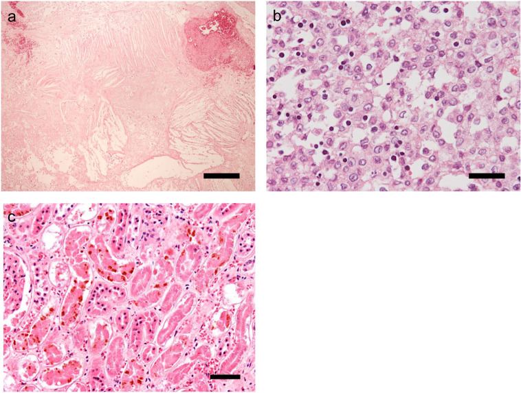
Fig. 4.
Histological examination by partial necropsy of the tumor tissue (a and b) and kidney tissue (c) with hematoxylin and eosin stain. a. Necrosis, hemorrhage, and cholesterin crystal depletion are observed over a wide area of the tumor (scale bar: 300 µm). b. Neoplastic cells with large nuclei and eosinophilic, granular cytoplasm proliferate diffusely and are accompanied by lymphocyte infiltration (scale bar: 30 µm). c. Extensive tubular necrosis and hemosiderin deposition in tubular epithelial cells are observed (scale bar: 50 µm).

