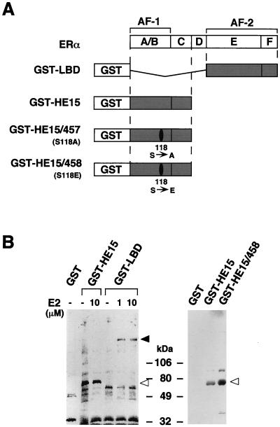
FIG. 1.
Detection of binding proteins to the hERα A/B domain in MCF-7 cells. (A) The hERα (with regions A to F shown) and the GST-ER fusion proteins used. (B) Binding proteins to the hERα A/B domain in MCF-7 cells. Aliquots of the 35S-labeled MCF-7 nuclear extract were incubated with glutathione-Sepharose beads loaded with GST alone, GST-HE15, GST-HE15/458, GST-HE15/457, or GST-LBD in the absence or presence of E2 at 1 and 10 μM. The bound proteins were subjected to SDS-PAGE (5 to 20% polyacrylamide gradient gel) followed by autoradiography. Open arrowheads indicate the position of a protein of 68 kDa. Size markers are indicated in kilodaltons. The solid arrowhead indicates the position of the SRC-1/TIF2 160-kDa family proteins (9, 14).

