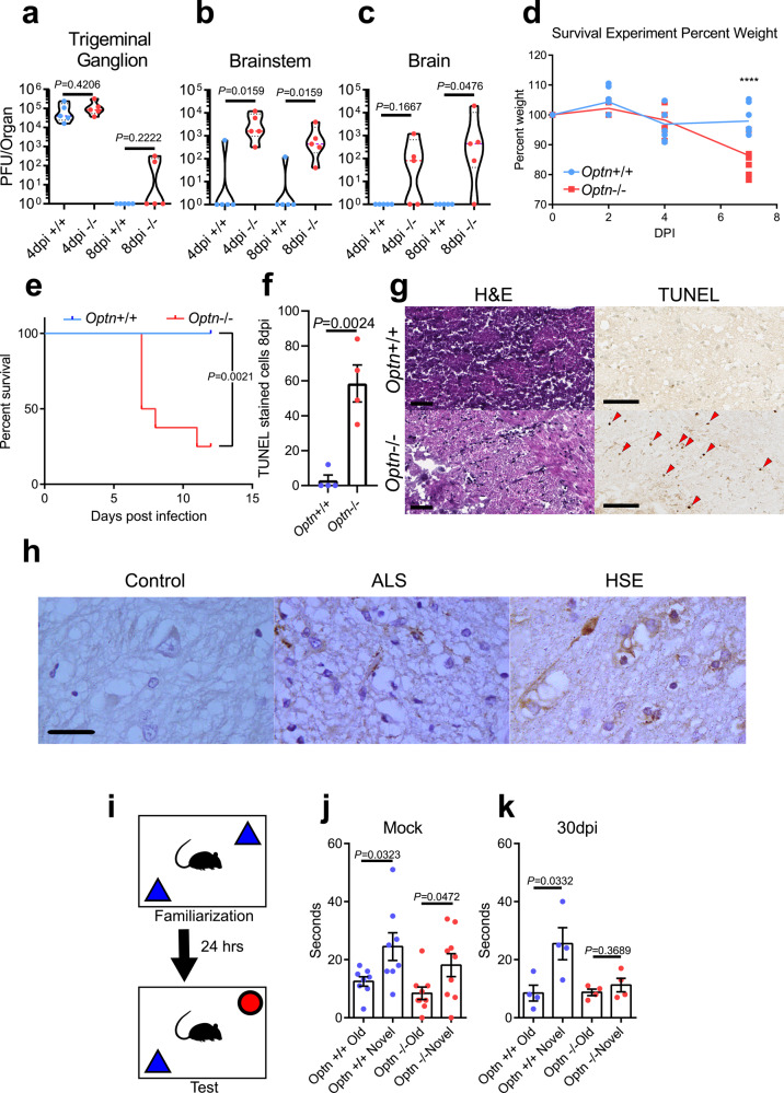
Fig. 5. OPTN is neuroprotective against herpes simplex virus encephalitis.
Mice were infected using corneal scarification using 5 × 10⁵ PFU of HSV-1 McKrae strain. a–c Tissue plaque assay for trigeminal ganglion (left), brainstem (middle), and brain (right) (n = 5 mice per group). Two-tailed Mann–Whitney U test was used where p < 0.05 indicates significance. d Weight changes of mice (n = 5 mice per group) up to 7dpi. Two-way repeated-measures ANOVA F(3,42) = 9.908, p < 0.0001. e Kaplan–Meier curve for HSV-1 infected Optn+/+ (n = 8) and −/− (n = 8) mice. Logrank test was used to compare curves with p < 0.05 being significant. f, g (left) Quantitation of TUNEL stained cells in brainstems of Optn+/+ and −/− mice 8dpi (n = 4 mice per group). (right) Representative images of H&E staining and TUNEL staining (dark brown) for brainstems. Arrows highlight TUNEL positive cells. Scale bar is 50 µm. h Immunohistochemistry staining of OPTN (dark brown) in human brainstem tissue from a healthy, amyotrophic lateral sclerosis (ALS), or HSE patient shows aggregation of OPTN protein expression in diseased tissues. Scale bar is 100 µm. i Schematic of Novel Object Recognition (NOR) test for mice. j mock-infected Optn+/+ and −/− mouse exploration times for old and novel objects. (n = 8 mice per group) (k) Optn+/+ and −/− mouse exploration times for old and novel objects at 30 dpi with 1 × 10⁵ PFU of HSV-1 McKrae strain (n = 4 mice per group). Data are presented as mean values ± SEM (f, j, k). Unless otherwise indicated, Two-tailed Student’s t test was performed for statistical analysis (α = 0.05). ∗p < 0.05; ∗∗p < 0.01; ∗∗∗∗p < 0.0001, ns not significant. Source data are provided as a Source Data file.

