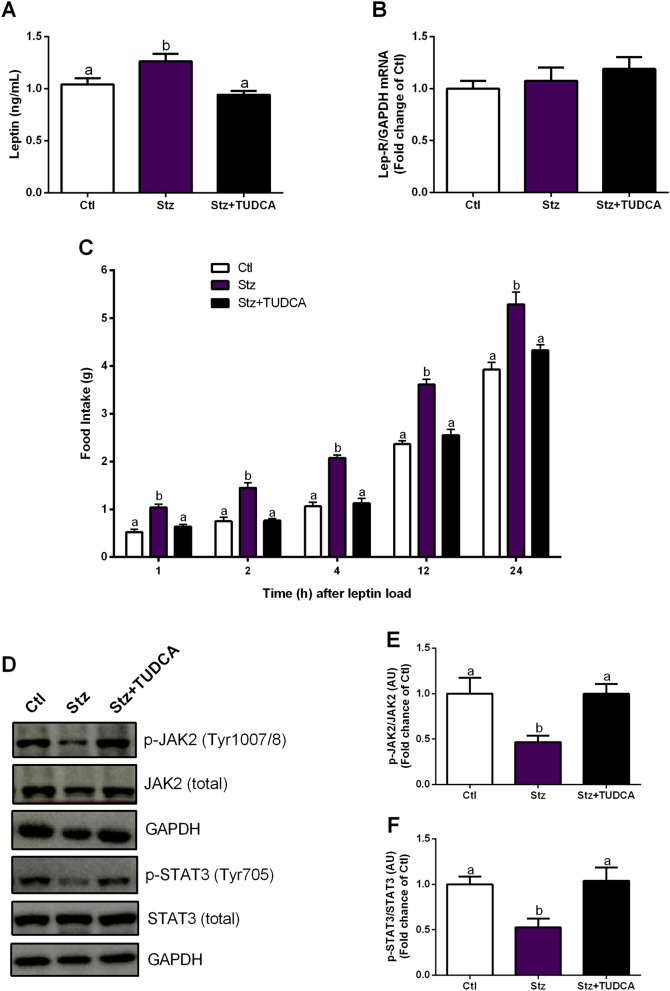
Figure 4.
Mice treated with TUDCA display ameliorated hypothalamic leptin signaling. Serum leptin levels (A) and gene expression of Lep-R (B) normalized by GAPDH in the hypothalamus. Acute food intake after leptin (5 mg/kg, i.p.) load (C). Representative western blot images of p-JAK2, total JAK2, p-STAT3, total STAT3 and GAPDH in the hypothalamus (D). Protein content of p-JAK2 (Tyr1007/8) normalized by total JAK2 (E) and p-STAT3 (Tyr705) normalized by total STAT3 (F) in the hypothalamus, 45 min after leptin (5 mg/kg, i.p.) load. GAPDH was employed as a housekeeping protein for total JAK2 and STAT3 normalization. Data are expressed as means ± SEM (n = 4–8). Different letters indicate significant differences between groups (P ≤ 0.05), based on one-way ANOVA test followed by Tukey post-hoc-test. AU arbitrary units.

