Figure 4.
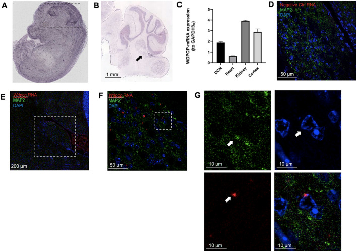
Wdpcp is expressed in the murine dorsal cochlear nucleus. In situ hybridization in an (A) 14.5 days post-conception mouse and (B) a C57BL/6 J mouse at P56 shows moderate expression of Wdpcp within the brain and DCN compared to surrounding tissues. The embryonic brain is demarcated by black dotted lines, while the DCN in the adult in situ data is marked by a black arrow. Quantification of Wdpcp RNA expression in a 2-month-old mouse shows relatively equivalent amounts in both the dorsal cochlear nucleus and cortex (C). Immunofluorescent imaging in 3-month-old mice combined with RNAScope shows colocalization of Wdpcp RNA in neurons (n = 5). Negative controls treated with a zebrafish slc17a8 probe at 40 × magnification (D). The DCN is outlined in white in the 10X panel (E). Images at 63 × show distinct Wdpcp RNA puncta (F). Enlargements of the space enclosed in the white box in panel F show colocalization of Wdpcp RNA in nuclei surrounded by MAP2 (G). White arrows denote location of MAP2 expression, nucleus of interest, and Wdpcp RNA puncta in split channels. Image credit: EMAGE Project (EMAGE: 18,852), and Allen Institute (https://mouse.brain-map.org/experiment/show?id=69012956).

