Figure 6.

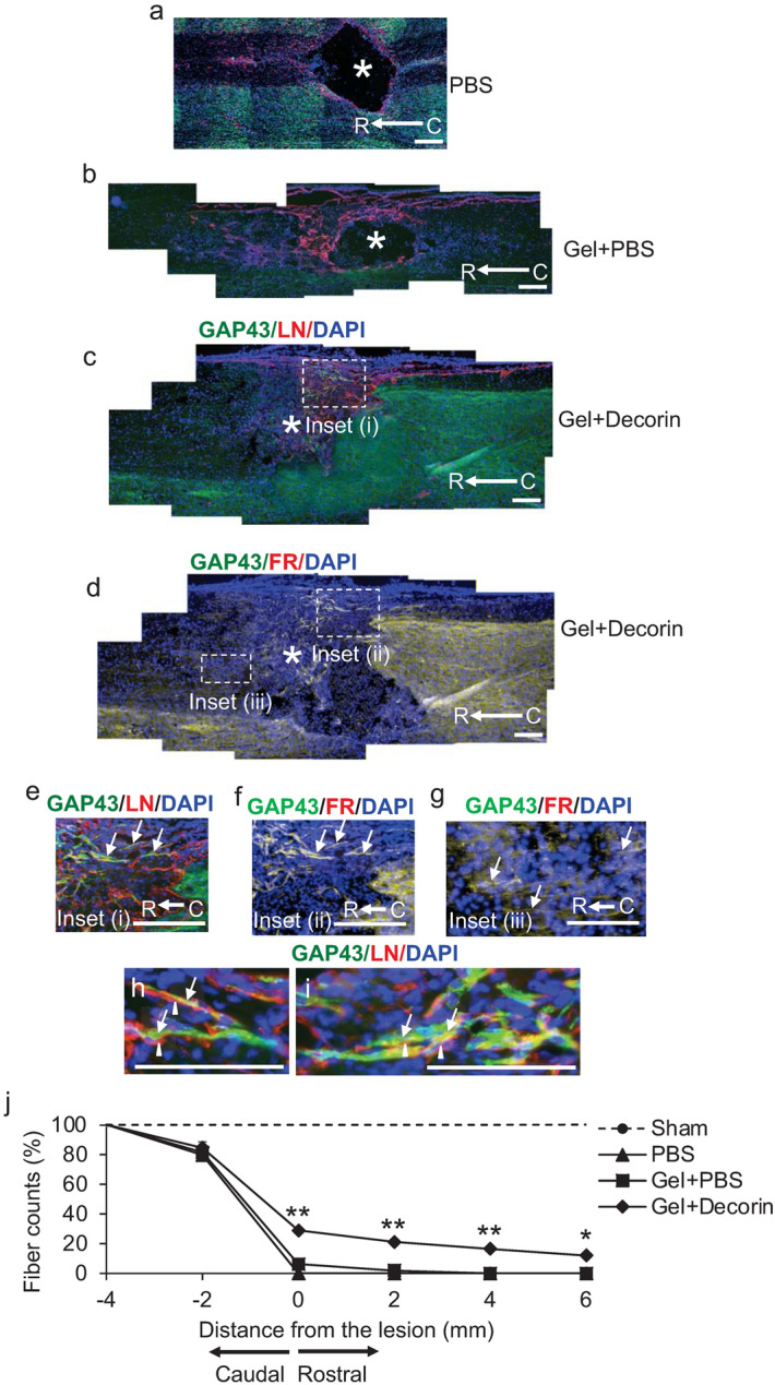
Gel + Decorin promotes DC axon regeneration. Little or no GAP43+ (green) axons were localised in (a) PBS or (b) Gel + PBS injected wounds with a cavity, devoid on LN+ immunoreactivity (red), remaining in the lesion core (*). DAPI = nuclear stain (blue). (c) No cavity was present in Gel + Decorin injected wounds, with many GAP43+ axons (green) traversing the lesion site, which was filled with LN+ (red) ECM fibres. (d) Merged image from an adjacent section from Gel + Decorin-treated animals costained for FR, GAP43 and DAPI to show overlap of FR and GAP43 staining in the same section. (e) High power view of boxed region in (c) which is labelled Inset (i) showing GAP43+ axons (arrows) regenerating through the lesion site from the caudal (C) to rostral (R) regions of the spinal cord (direction of regeneration is C to R). (f) High power view of same boxed region in (c), labelled Inset (ii), which was co-labelled with the bidirectional axon tracer, FluoroRuby (FR), demonstrating overlap of GAP43+ with FR+ axons (arrows). (g) High power view of boxed region in (c) which is labelled Inset (iii), showing FR+ axons (arrows) that have regenerated into the rostral spinal cord. (h) and (i) represent high power views GAP43+ regenerating axons (arrows), traversing along LN+ fibres (arrowheads) within the lesion site of Gel + Decorin-treated animals. (j) Quantification of GAP43+ axons at different distances from the caudal through the lesion site and in the rostral spinal cord. Scale bars in (a–h) = 200 µm. ** = P < 0.001, * = P < 0.05, one-way ANOVA with Dunnett’s post hoc test. n = 6 rats/group, 3 independent repeats, total n = 18 rats/group.
