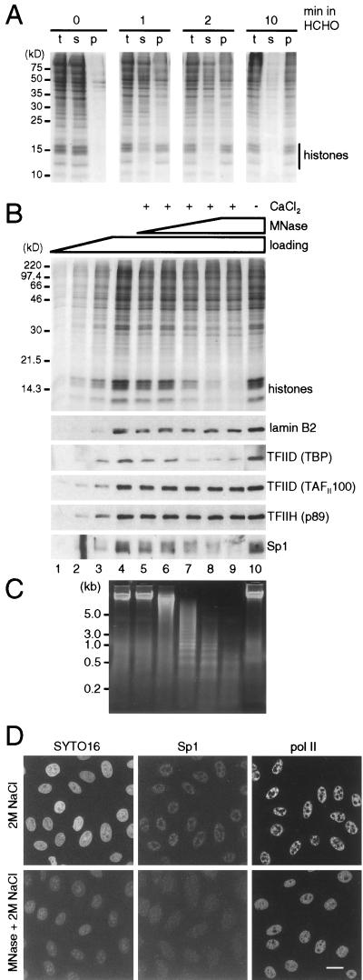
FIG. 4.
Protein complexes detected after cross-linking with formaldehyde. (A) Establishment of conditions for cross-linking. Cells were either untreated or treated with 1% formaldehyde for 1, 2, or 10 min and then with 0.1% formaldehyde for 3 min, extracted with 2 M NaCl, and centrifuged. Total (t) proteins in extracted cells and those in supernatant (s) and pellet (p) were resolved in a 15% gel and stained with Coomassie blue. After 0, 1, 2, and 10 min in 1% formaldehyde, 0, 65, 80, and 95%, respectively, of the histones were recovered in the pellet. (B) Proteins remaining after treatment of fixed cells with micrococcal nuclease (MNase). Cells were fixed with 1% formaldehyde for 2 min and then with 0.1% formaldehyde for 3 min, lysed with saponin, incubated with or without micrococcal nuclease ± CaCl2 at 37°C, chilled on ice, extracted with 2 M NaCl, and pelleted; then the proteins in the pellet were resolved on a gel, stained with Coomassie blue (top), or blotted and probed with antibodies directed against the proteins indicated. Selected regions of the blots are shown below. Lanes 1 to 4: 1/8×, 1/4×, 1/2×, and 1× loading of cells treated similarly but incubated on ice; lanes 5 to 9: samples incubated with 0, 0.0016, 0.008, 0.04, or 0.2 U of MNase per ml and 1 mM CaCl2; lane 10, sample incubated with 0.2 U of MNase per ml without CaCl2. (C) Photograph of DNA fragments from fixed cells treated with micrococcal nuclease. DNA fragments in samples 4 to 10 in panel B were run on an agarose gel and stained with ethidium. (D) Micrographs illustrating the distribution of Sp1 and Pol II. Cells on coverslips were fixed, treated with micrococcal nuclease or left untreated (as in panel B, lanes 4 and 9), extracted with 2 M NaCl, and refixed with 4% formaldehyde. Then Sp1 and the largest subunit of Pol II were indirectly immunolabelled with Cy3, nucleic acids were counterstained with SYTO16, and equatorial sections through cells were obtained under a confocal microscope. Both Sp1 and Pol II survive extraction with 2 M NaCl and are found in many small foci throughout the nucleoplasm but not in nucleoli (top row). After nuclease treatment, considerable amounts of nucleic acids and Sp1 are extracted, but most of the Pol II remains. Bar, 20 μm.

