Fig. 2.

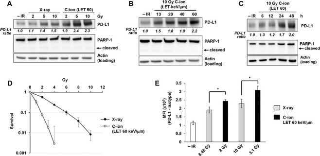
Carbon-ion irradiation upregulates PD-L1 protein expression in U2OS cells. (A) PD-L1 protein expression after carbon-ion irradiation with LET at 60 keV/μm was higher than that detected after X-ray irradiation. U2OS cells were harvested at 48 h after exposure to 2, 5, or 10 Gy X-ray or carbon-ion irradiation. Poly(ADP-ribose) polymerase (PARP-1) cleavage was examined to confirm that apoptosis was not induced in the analyzed cells. (B) PD-L1 upregulation in U2OS cells was examined 48 h after irradiation with 10 Gy of carbon-ions with LET at 13, 20, 40 and 60 keV/μm. (C) PD-L1 upregulation in U2OS cells was examined at the indicated time points after irradiation with 10 Gy of carbon-ions with LET at 60 keV/μm. (D) A colony formation assay was performed in U2OS cells to calculate RBE comparing X-ray and carbon-ion with LET at 60 keV/μm. (E) Flow cytometry analyses for cell-surface PD-L1 were performed in U2OS cells 48 h after 6.48 Gy or 10 Gy X-ray vs. 2 or 3.1 Gy carbon-ion irradiation with LET at 60 keV/μm, to set a similar RBE dose. The statistical significance following Bonferroni’s correction is shown. *P < 0.0125. In A–D, the signal intensities of PD-L1 and actin were measured using ImageJ. The PD-L1 signal was normalized to that of actin; subsequently, the ratio of PD-L1 upregulation was normalized to that detected in non-irradiated cells.
