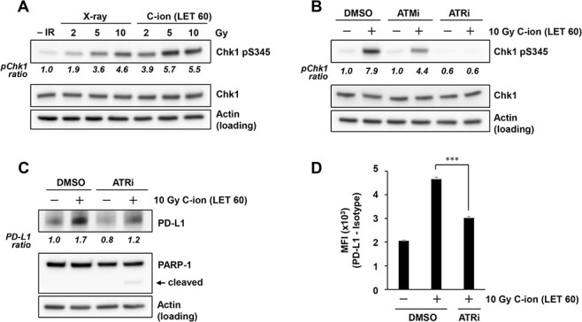
Fig. 3.

Carbon-ion irradiation upregulates PD-L1 expression in an ATR-dependent manner. (A) Chk1 phosphorylation at S345 in U2OS cells was examined at 2 h after 2, 5 or 10 Gy X-ray or carbon-ion irradiation with LET at 60 keV/μm. Because resection and Chk1 phosphorylation peak at ~2 h after irradiation, Chk1 phosphorylation was examined at 2 h after exposure to IR. (B) Chk1 phosphorylation at S345 in U2OS cells was examined in the presence of 10 μM ATM (KU55933) or 10 μM ATR inhibitor (VE821) at 2 h after irradiation with 10 Gy of carbon-ions with LET at 60 keV/μm. (C) PD-L1 expression in U2OS cells was examined in the presence of 10 μM ATR inhibitor (VE821) at 48 h after 10 Gy carbon-ion irradiation with LET at 60 keV/μm. (D) Cell surface PD-L1 expression in U2OS cells was examined in the presence of 10 μM ATR inhibitor (VE821) at 48 h after irradiation with 10 Gy of carbon-ions with LET at 60 keV/μm. The statistical significance following Bonferroni’s correction is shown. ***P < 0.0005. In A–B, the signal intensities of Chk1 pS345 and Chk1 were measured using ImageJ. The Chk1 pS345 signal was normalized to that of Chk1; subsequently, the ratio of Chk1 pS345 upregulation was normalized to that detected in non-irradiated cells. PD-L1 quantification in C was performed as described in Fig. 1.
