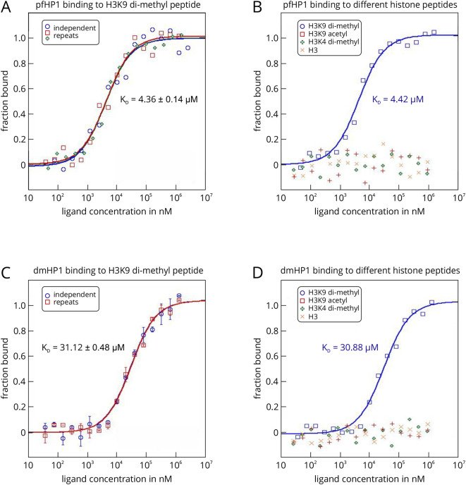
Figure 4. MST analysis of the interactions between P. falciparum and D. melanogaster HP1 and different histone peptides using the label-free MST technology.
The HP1 proteins from P. falciparum (pfHP1) and D. melanogaster (dmHP1) served as the targets in this example. In this example, it was not necessary to attach a fluorescent label to the targets, because both proteins featured high enough molar extinction coefficients to analyze the interactions on a Monolith NT.LabelFree device. A prerequisite for this was also that the histone peptides did not contain tryptophan and tyrosine residues (no fluorescence above 300 nm).

