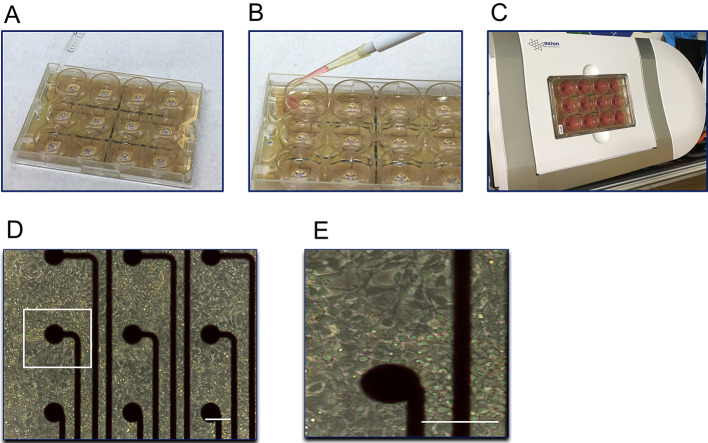
Figure 2. Representative images of MEA plate handling.
MEA plate with sterile water surrounding the wells to prevent the evaporation (A). Recommended method of pipetting medium into MEA wells to avoid damaging the cells (B). MEA plate placed in the plate cavity (C). hiPSC-derived neurons plated around recording electrodes (D). A zoomed-in view of the marked white square in panel (D), showing neurons surrounding one of the recording electrodes (E). Scale bars = 100 μm.

