
Fig. 3.
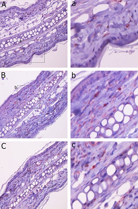
VEGF expression was observed in the ear skin of C3H mice using immunohistochemistry. VEGF expression was found in scattered fibroblasts in the dermis. A: control group. B: 2 Gy group. C: 15 Gy group. a, b and c: Enlarged image of positive cells for A, B and C, respectively. The number of VEGF-positive cells at 24 h following 2 Gy irradiation (++) slightly increased compared to that in the control group (+); however, the expression of VEGF in the 15 Gy (+) irradiation group revealed no difference compared to that in the control group (+).