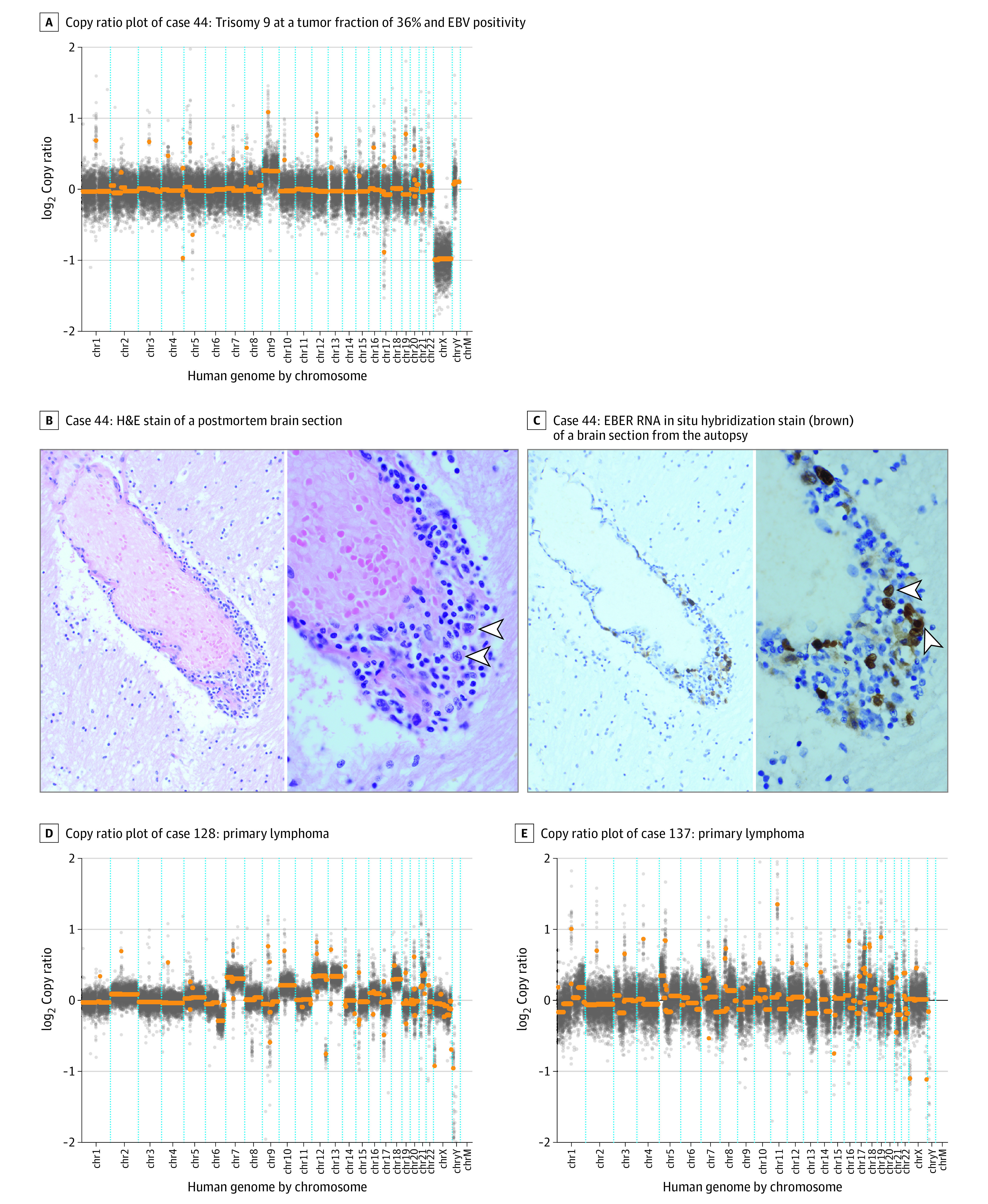
Figure 2. Undiagnosed Primary CNS Lymphoma Cases.

A, Conventional methods of brain biopsy and cytologic testing of the cerebrospinal fluid (CSF) were nondiagnostic in case 44. Flow cytometry showed atypical cells and was not definitive. Diagnosis was confirmed by autopsy 2 weeks after the CSF sampling. B, Arrowheads highlight large, perivascular B cells that have large, irregular nuclei with loose chromatin; hematoxylin-eosin (H&E) stain was used, with original magnification ×200 (left image) and ×1000 (right image). C, The large cells were stained positive (brown) for Epstein-Barr encoding region (EBER) in situ hybridization as evidence of Epstein-Barr virus (EBV) RNA expression in malignant lymphoma. D and E, Copy ratio plots are from 2 patients enrolled in the neuroinflammatory disease case-control study.
