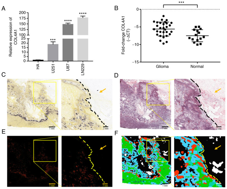
Figure 2.
Expression levels of COL4A1 in glioma are abnormally increased. (A) The expression levels of COL4A1 in glioma cell lines were significantly higher than those in astrocytes based on RT-qPCR detection. (B) COL4A1 expression levels in glioma tissue samples were significantly higher than those in normal brain tissue according to RT-qPCR detection. ***P<0.001; ****P<0.0001. (C-F) COL4A1 detection in samples from the Ivy Glioblastoma Atlas database. Scale bars, 1 mm. (C) Histological section of glioblastoma under the light microscope. (D) Histological section of glioblastoma and adjacent tissue stained with H&E. (E) Detection of COL4A1 expression levels in tumor and paratumoral tissue by in situ hybridization. (F) Tumor feature annotation of histological section of glioblastoma and adjacent tissue; Green, Cellular Tumor; Black, non-tumor cell infiltration area; Red, Area of vascular proliferation; Blue, Perinecrotic zone; Gray-green and purple, Pseudopalisading cells. The yellow arrows represent non-neoplastic infiltrates. COL4A1, collagen α-1 (IV) chain; RT-qPCR, reverse transcription-quantitative PCR; HA, human-derived astrocytes.

