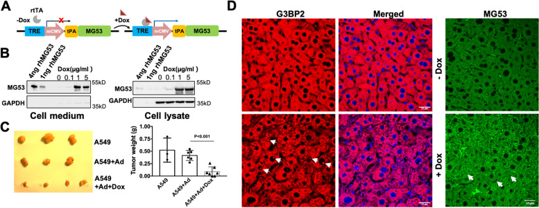
Fig. 7.
Overexpression of MG53 suppresses A549 xenograft tumor growth in vivo. A Schematic representation of the Ad-tPA-MG53 cDNA under the control of a tetracycline response element (TRE) [60], allowing to tailor the expression and secretion of MG53 in a Dox-inducible manner. B A549 cells were infected with adenovirus (Ad-TRE-tPA-MG53), induced with or without Dox for 24 hr, then cells and conditioned culture medium were harvested and analyzed. Western blot analysis revealed that with addition of Dox (0.1, 1, and 5 μg/ml), dose-dependent elevation of MG53 (right panel) with concurrent secretion of MG53 into the culture medium was observed. GAPDH served as a loading control. C A549 xenograft weight from mice treated with Dox were significantly smaller than those from animals treated with the control saline group or non-Dox treatment group (n>6 for each group). D Primary tumors from mice were subjected to IHC staining with anti-G3BP2 antibody. The nucleus was stained with DAPI (blue). Arrowheads indicate the nuclear localization of G3BP2. E IHC staining with MG53 showed increased levels of MG53 with Dox-treatment (bottom panel). Arrowheads indicate nuclear localization of MG53. Because antibodies against G3BP2 and MG53 used for the immunostaining were all derived from rabbits, separate IHCs with G3BP2 and MG53 were conducted with different sections of the xenograft

