
Fig. 2.
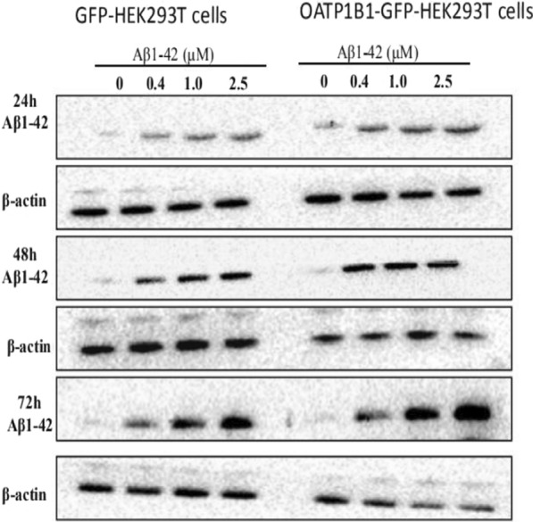
GFP-HEK293T cells and OATP1B1-GFP-HEK293T cells were cultured in 6-well plates at 2.5 × 10⁵ cells/well and after adherence, were treated withAβ1-42 (0, 0.4, 1, 2.5 μM) for 24, 48, and 72 h. Following treatment, Aβ1-42 was determined in the cell lysates by western blotting (WB). Compared to the GFP-HEK293T cells group, the uptake of Aβ1-42 protein in the OATP1B1-GFP-HEK293T group increased with the increase in Aβ1-42 concentration. The increase was significant with the increase in incubation time. Aβ1-42 bands to oligomer was about 64KD. β-actin served as an internal control