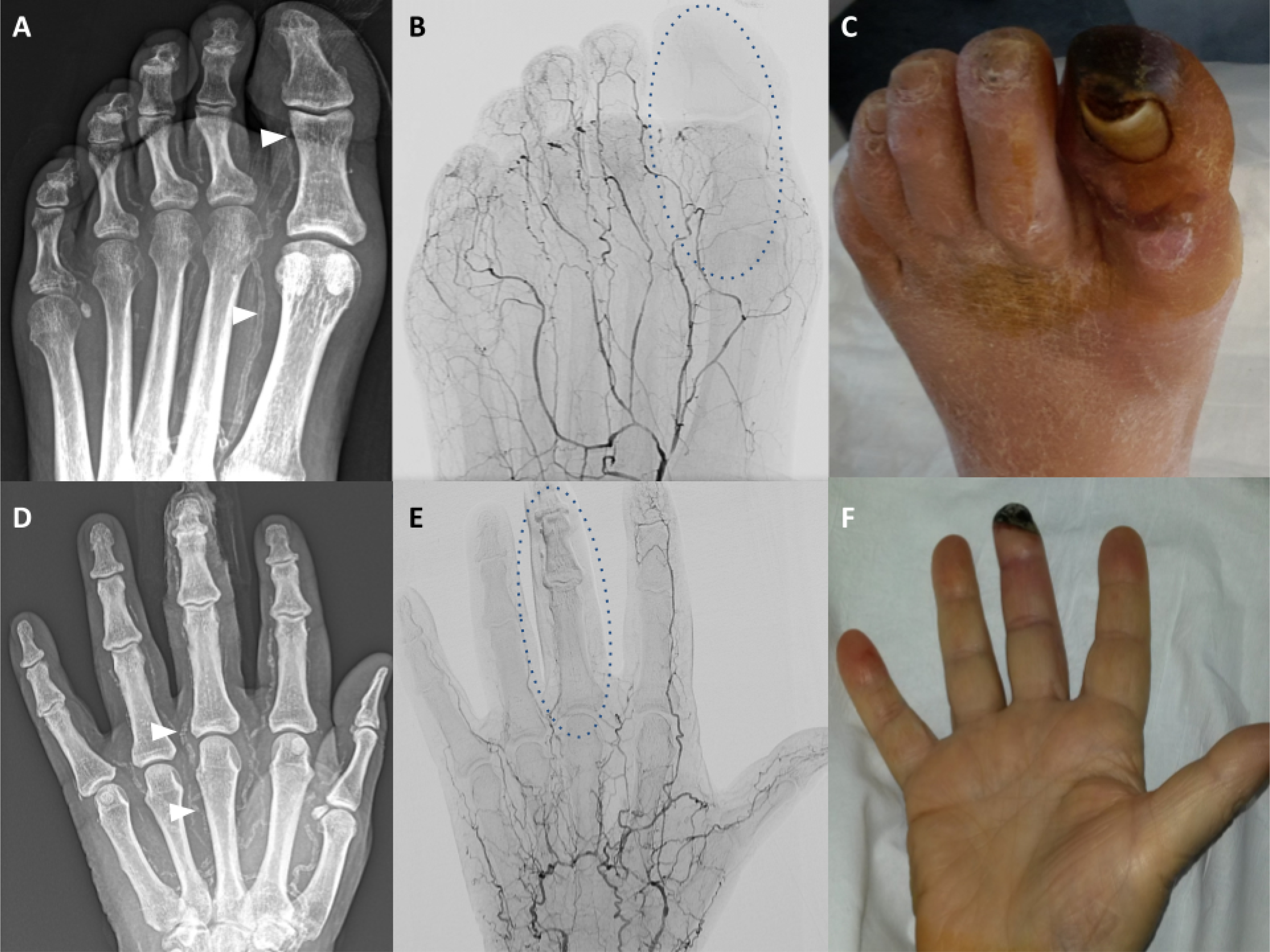
Figure 8. MAC in limb arteries.

Male with the gangrene of the great toe left. (A) Native X-ray image of the left foot; heavy calcifications of the metatarsal and toe arteries, particularly of the first metatarsal and great toe arteries are shown (white arrowheads). (B) Angiographic X-ray image corresponding to A; shown is the extensive destruction of the small arteries; paucity of the distal arterial supply is highlighted in the context of the great toe (oval shaped broken line). (C) Photograph of the left foot corresponding to A; shown is extensive necrosis of the left great toe. Male with the gangrene of the mid-finger left. (D) Native X-ray image of the left hand; heavy calcifications of the metacarpal and finger arteries are shown (white arrowheads). (E) Angiographic X-ray image corresponding to A; shown are multiple occlusions of the metacarpal and finger arteries and virtual absence of arterial blood flow to the fingers (exemplified by the mid-finger, oval shaped broken line). (F) Photograph of the left hand corresponding to A; shown is distal necrosis of the mid-finger left.
