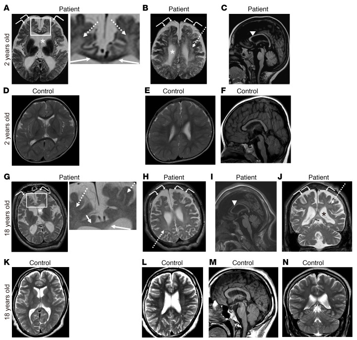
Figure 1. Brain MRI.
Brain MRI of the patient (A–C and G–J) and age-matched normal controls (D–F and K–N) at 2 years (A–F) and 18 years (G–N) of age. T2-weighted axial (A, B, D, E, G, H, K, and L), T2-weighted coronal (J and N), and T1-weighted sagittal (C, F, I, and M) images are shown. The boxed areas in A and G were enlarged and are shown on the right. Cerebral atrophy with ventriculomegaly (asterisks) and an enlarged subarachnoid space (curved arrows) were observed (A, B, G, H, and J). An abnormal T2 hyperintense signal of the diffuse cerebral white matter (dashed arrows) indicated hypomyelination (A, B, G, H, and J). The patient’s corpus callosum at 2 years of age showed a T2 hypointense signal (arrows, magnified field in A), indicating normal myelination, while an abnormal T2 hyperintense signal in the corpus callosum (arrows, magnified field in G) was observed in the patient at 18 years of age. Midline sagittal T1-weighted brain MRI demonstrated a thin corpus callosum (arrowheads in C and I). Cerebral atrophy did not appear to have progressed in the patient between the ages of 2 and 18 years.

