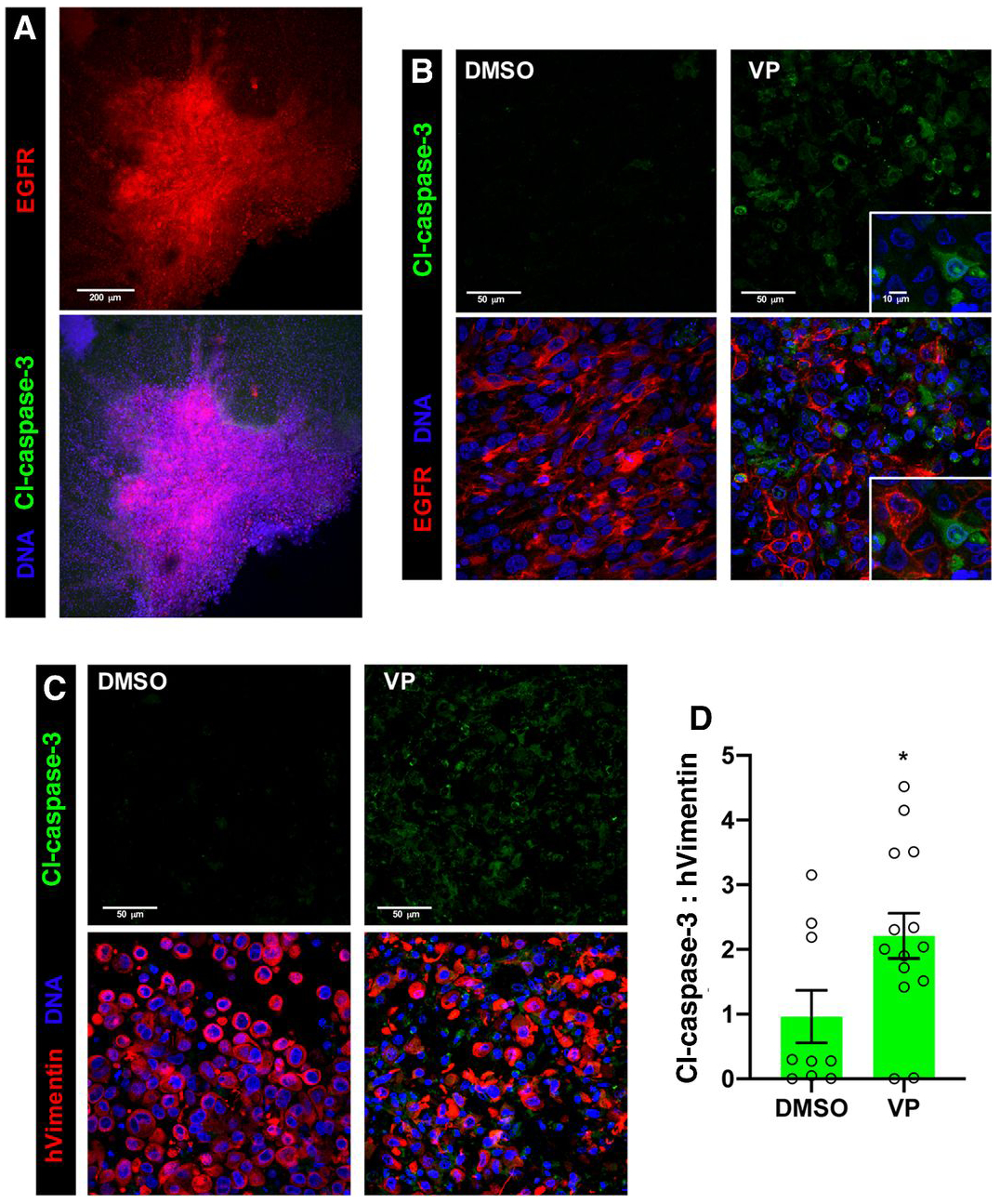
Figure 4. Verteporfin treatment induces apoptosis in GBM cells cultured in ex vivo organotypic cultures.

Ex vivo organotypic slice cultures created from GBM39 xenografts implanted into NSG mice, treated with DMSO (control), 1μg/mL verteporfin (VP) for 24 hours, and/or other indicated agents, and then stained and imaged with confocal microscopy. DRAQ7 DNA dye (blue) labels all cell nuclei in tumor and stroma. 3 μm optical projections.
(A) Low magnification view of a representative control slice culture showing tumor integrated into surrounding normal brain tissue, tumor cells marked by EGFR immunostaining (red); cleaved-Caspase-3 immunostaining (green) in DMSO treated tumor and normal tissue.
(B) Immunostaining for EGFR (red), with verteporfin treatment. Inset shows close-up of apoptotic cells (green, cl-Caspase-3) adjacent to EGFR-positive tumor cells.
(C) Immunostaining for cleaved-Caspase-3 (Cl-Caspase-3, green, upper panels), labeling apoptotic cells, and human Vimentin (hVimentin, red, lower panels overlay), which specifically labels human tumor cells in the mouse brain parenchyma in organotypic tumor xenograft slice cultures.
(D) Ratios for total cleaved-Caspase-3 and hVimentin immunostaining in organotypic slice cultures; values measured in 30-50 μm confocal z-stacks using Imaris. Graphs shows values from individual experimental samples from multiple experiments.
*p≤.05 with unpaired t test.
