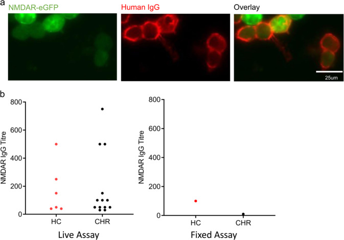
Fig. 2. NMDAR IgG seroprevalence and associations of seropositivity—antibodies detected using live assay.
a Representative example of NMDAR IgG-positive live CBA. Human IgG is labelled red using a fluorescent secondary antibody (AlexaFluor 568 goat anti-mouse IgG (H + L); Invitrogen) and shown binding to cells that co-express eGFP (green). Scalebar = 25 µm. b Distribution of NMDAR IgG titre by group and assay type (fixed vs. live assay).

