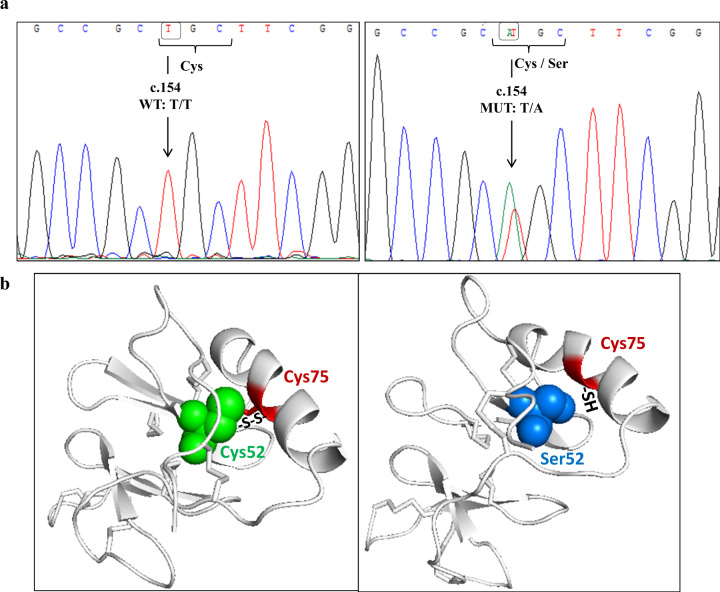
Fig. 2.
a AVP gene’s exon 2 chromatogram for two patients. On the left: wild-type sequence with T allele at position c.154; On the right: mutant heterozygous sequence with the mutation at position c.154 (T → A) that leads to an amino acid substitution from cysteine (Cys) to serine (Ser). b Three-dimensional structure of AVP-NPII protein. On the left: Wild-type protein with cysteine at position 52 (Cys52) (represented as green spheres) that forms disulfide bridge (S-S) with cysteine at position 75 (Cys75) (depicted as red region). On the right: mutant protein with serine at position 52 (Ser52) that prevents the formation of disulfide bond with the sulfhydryl groups (-SH) of cysteine at position 75 (Cys75). Ser52 and Cys75 are represented as blue spheres and red sticks in helix region, respectively

